Pet Dentistry
Interesting Dental Cases
Prevention and treatment of periodontal disease, abscessed teeth, and cavities keep pets comfortable. These problems all cause pain in animals just as they do in people. Pets also can suffer from oral tumors, malpositioned teeth, and other diseases of the mouth.
Retained Canine Teeth
Adult Teeth That Did Not Erupt Properly
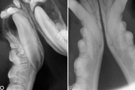
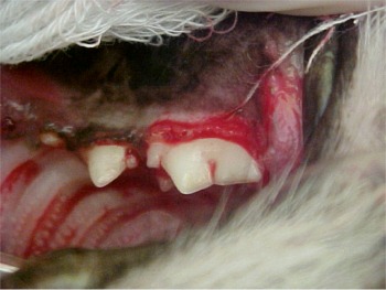
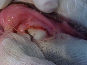
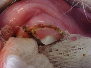
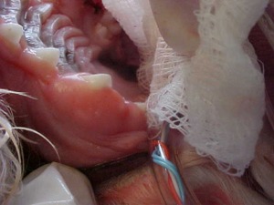
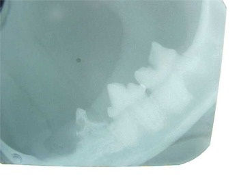
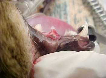
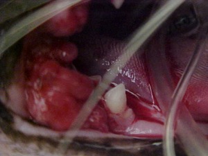
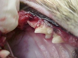
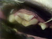
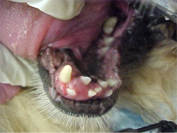
This little cutie is Violet. She is a 7-month-old Boston terrier. Many of her adult teeth did not come in all the way, and her lower canine teeth didn’t erupt at all. When we spayed Violet at 6 months of age, all her adult teeth should have been in already, but the lower adult canines were nowhere to be seen, and her baby canine teeth were still present when they should have fallen out. Dr. Boss attempted to remove the baby canines to get them out of the way so the adult teeth could erupt properly.
We consulted with our local Grafton, WI veterinary dental specialist, Dr. Dale Kressin, about what to do. He told us that the cystic fluid building up where the malpositioned teeth should be would eat away at the jawbone and destroy it, so those retained teeth needed to be removed.
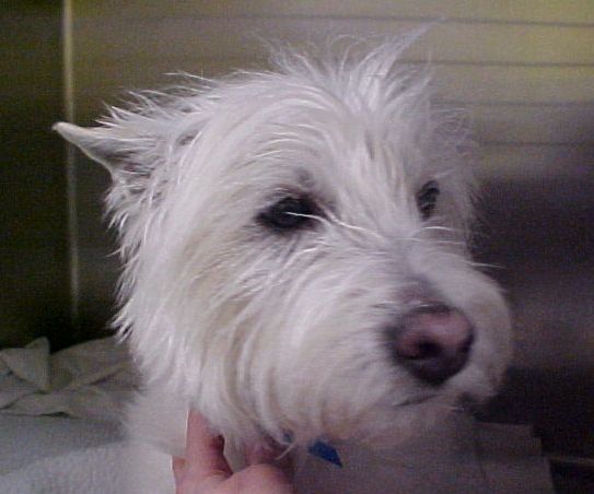
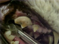
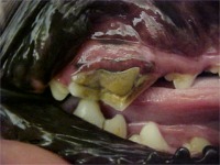
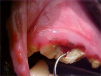
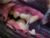
This is Weilly, who is a 9-year-old West Highland white terrier (Weilly the Westie!) He came in to Best Friends for a dental cleaning. Once we cleaned the plaque and tartar from the teeth we could see three problems. He had a 4th upper premolar tooth that was discolored, which signified that at least one root of this three rooted tooth was dead, leaving the tooth without its normal blood supply. In addition, both his lower 1st molars had holes in them on the tongue sides of the teeth (where they couldn’t be seen without anesthesia).
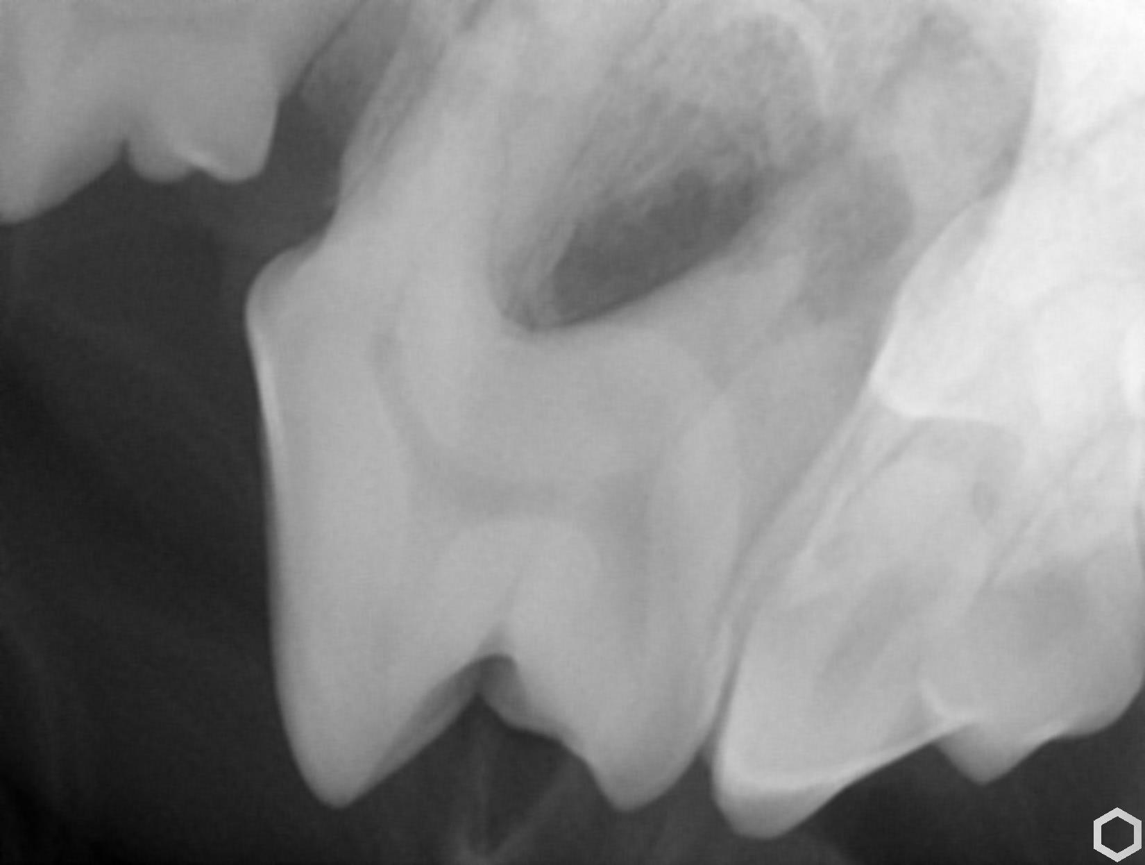
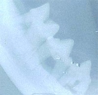
X-rays of these three teeth revealed holes in the enamel and dentin of the teeth. We call these cavities “resorptive lesions” because the tooth structure is being reabsorbed back into the body, leaving a gaping hole and exposed pulp and nerve tissue. Ouch! The large, irregularly shaped hole in the upper 4th premolar had almost eaten entirely through the bone between the two roots and one of the roots as well.
Weilly’s owners were faced with the choice between extracting all three teeth, for a good sum of money, or extracting just the upper tooth and having root canals and fillings for the lower molars by a dental specialist, for an even greater sum of money – several thousand dollars. These are three of the four largest chewing teeth in the dog’s mouth, so saving two of them would be desirable, owner’s financing permitting. Unfortunately for both the pet and the pet owner, Weilly is likely to develop more resorptive lesions in the future, as the cause is unknown and there is no preventative treatment that would safeguard the rest of his teeth. Which option would you choose if this were your pet?
Common Dental Problems
Below are some pictures of common problems we see.
Cat, moderate tartar and periodontal disease, before dental cleaning and after dental cleaning.
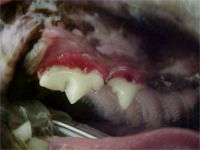
This puppy’s teeth did not erupt normally through the gums, and the teeth were covered with excess gum tissue. We used a CO2 laser to remove the excess gum tissue so she could eat and chew normally.
This cat has a cavity called a Feline Oral Resorptive Lesion or FORL. It has eaten away the crown of the tooth. The dental x-ray shows the same tooth. These lesions are very painful, and affected teeth should be removed.
About 40% of cats develop one or more of these cavities, usually between the ages of 2 and 10 years. If one cavity develops, there is a 75% chance that additional teeth will also develop them.
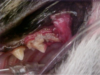
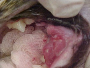
Stomatitis means inflammation in the mouth. This is usually a problem for cats. It is thought that affected cats have developed an allergy to the plaque bacteria in their mouths. The allergic reaction causes the gums and mouth tissues to become red, swollen and sore. Many times there are also cavities and periodontal disease involved. Infection with the Feline Leukemia or Feline Immunodeficiency viruses can lead to gingivitis or stomatitis. The most successful treatment is to remove the teeth where the inflammation exists, although we may try antibiotics and anti-inflammatory medications first.
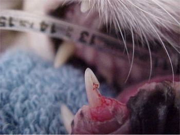
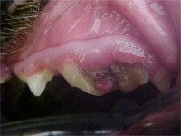
The root toward the left is no longer attached to the bone at all – this tooth is soon to fall out.
Periodontal disease occurs when infection works its way under the gums and along the tooth roots. First, the gums get red and sore, then the gum and bone start to recede, exposing more and more of the tooth roots.
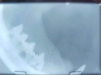
Eventually, there will be so little attachment of the root to the bone that the tooth will fall out. These X-rays show receded bone in the jaw of a cat and a dog.
The before and after dental cleaning views of a cat show what usually causes the periodontal disease (heavy tartar), and the partially exposed tooth roots after the teeth are cleaned.
Notice how red and sore the gums look.
For more in-depth explanations and photos of more unusual dental problems go to: dentalvet.com
To help prevent some of these problems, visit our Dental Cleaning page for a demonstration on cleaning of your pet’s teeth. We send our dental referrals to Animal Dental Center – Milwaukee and Oshkosh, WI.


