Epulides
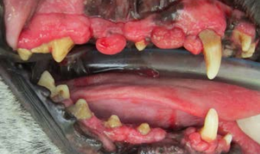
The most common type of tumor in the mouths of dogs is called an epulis. The plural of this latin-based name is epulides. Epulides most commonly arise in response to the accumulation of plaque and tartar on the teeth. Gum inflammation can lead to gum recession but it can also lead to lumps instead. These lumps usually hang over or cover up the teeth, which then leads to even more plaque and tartar build-up underneath. Epulides should be removed at the time of dental cleaning, and seeing them usually indicates periodontal disease.
We can see epulides in any dog but some dog breeds have much higher tendency to form epulides or to have hyperplastic (excessive) gum growth. Boxers, bulldogs and mastiffs are particularly prone. These dogs may need extensive gum surgery on a regular basis to keep this under control. Without removal, the overgrown gums tissue may bleed, cause pain from chewing and eventually lead to tooth loss.

In dogs, there are several types of epulides. The most common type, a fibromatous epulis, appears on a stalk of tissue, much like a mushroom, or as an unmoving lump. It is usually pink in color and has a smooth surface. It may appear as an enlargement on the gum tissue near an incisor, canine, or premolar tooth. These are the most common and least concerning type as they don’t invade into the bone.
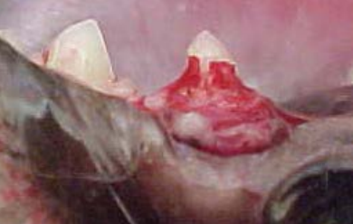
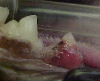
In cats, epulides often arise where there are cavities, called resorptive lesions, in the teeth. If part of a tooth is obscured by a gum lump in a cat, there may be plaque underneath but more likely there is a hole in the tooth. Resorptive lesions are painful and destroy the tooth as the crown is eaten away. They should be addressed promptly to avoid unnecessary pain. The picture to the left shows the gum covering a resorptive lesion. The right-hand picture shows the hole in crown of the tooth underneath the epulis.


